Publications
2025
- Impact Factor < 5
 Mature CD209+CD83+CCR7+ dendritic cells infiltrate the arterial wall in giant cell arteritis and derive from in-situ monocyte differentiationAndré Ramon, Hélène Greigert, Baptiste Lamarthée, Corentin Richard, Alexis Varin, Claudie Cladière, Coraline Genet, Marion Ciudad, Noémie Klopfenstein, Roman Praliaud, Guillaume Brenac, Georges Tarris, Laurent Martin, Louis Arnould, Pierre-Henry Gabrielle, Catherine Creuzot Garcher, Paul Ornetti, Sylvain Audia, Romain Boidot, Jean-Francis Maillefert, Bernard Bonnotte, and Maxime SamsonSci Rep, Oct 2025
This study aimed to characterize arterial dendritic cells (DCs) in polymyalgia rheumatica (PMR) and giant cell arteritis (GCA). Bulk RNA-sequencing, RT-PCR and immunofluorescence analyses were performed from temporal arteries from GCA, PMR and control patients. Public single-cell RNA-seq (scRNA-seq) data on Peripheral Blood Mononuclear Cells (PBMCs) were analyzed from three GCA and three control patients. Bulk RNA-Seq and RT-PCR analyses demonstrated a high level of expression of DC lineage markers (CD209), DC maturation markers (CD83, CCR7) and chemokines associated with DC maturation in GCA arteries. The level of expression of DC lineage and DC maturation associated genes was significantly lower in PMR than in GCA arteries and similar between PMR and control arteries. GCA arteries expressed high levels of GM-CSF and IFNG mRNA. ScRNA-seq analysis of GCA PBMCs demonstrated high expression of IFNGR and CSF2R by classical monocytes and cultures of CD14(+) monocytes with GM-CSF and IFN-γwere able to promote their differentiation into monocyte derived-DCs (mo-DCs). This work provides evidence that mo-DCs infiltrate GCA lesions and could be generated under the influence of GM-CSF and IFN-γfrom monocytes infiltrating the arterial wall. Mo-DCs could play an important role in GCA pathogenesis and be targeted by GM-CSF and/or IFN-γinhibitors.
- Preprint
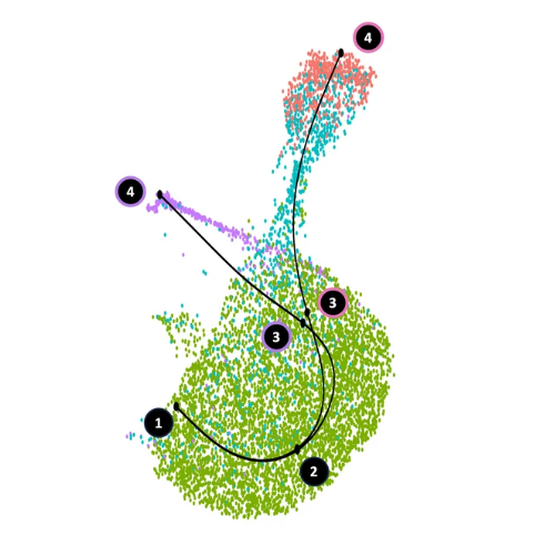
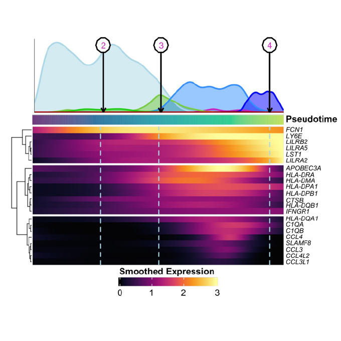 A subset of pro-inflammatory CXCL10+ LILRB2+ macrophages derives from recipient monocytes and drives renal allograft rejectionAlexis Varin*, Jovanne Palvair*, Lennie Messager*, Jamal Bamoulid, Yacine Benchikh, Jasper Callemeyn, Mélanie Chaintreuil, Ludivine Dal Zuffo, Didier Ducloux, Imane Farhat, Mathieu Legendre, Laurent Martin, Florian Renosi, Xavier Roussel, Thibaut Vaulet, Maarten Naesens, Claire Tinel, and Baptiste LamarthéemedRxiv, Apr 2025
In solid organ transplantation, monocytes and macrophages play a cross-cutting role in the rejection process, irrespective of the transplanted tissue and the type of rejection. Here, we integrated multiple single-cell assays (>150,000 cells) with a broad spectrum of blood-derived and renal allograft-derived cells. We observed 6 myeloid cell trajectories enriched in the allograft during rejection, ranging from circulating CD14+ monocytes to differentiated macrophages in the kidney, with one trajectory culminating in a pro-inflammatory macrophage expressing CXCL9 and CXCL10. By analyzing over 850 biopsies using deconvolution, we report that they are absent in pre-transplant allografts, while these CXCL10+ macrophages are the immune cells most associated with inflammation during acute rejection. Furthermore, a survival study of over 500 biopsies indicates that they increase the risk of graft loss independently of other immune cells. CXCL10+ macrophages differentiate from recipient monocytes, and we have identified 6 major genes associated with their differentiation, including LILRB2. In vitro, mimicking allogenic activation of blood monocytes via the CD47/SIRP-a axis induced overexpression of LILRB2, suggesting that CXCL10+ macrophages are activated by this pathway. Finally, we show that macrophages overexpressing LILRB2 induce the proliferation of autologous T lymphocytes. Altogether, the present study provides further insight into the pro-inflammatory axes of recipient-derived monocytes/macrophages, and suggests LILRB2 as a therapeutic target.